Clinical Insights
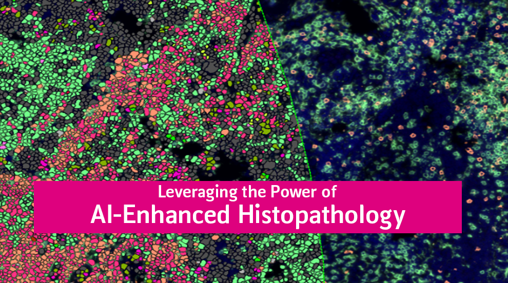
Power Your Oncology & Immunology Trials with AI-Enhanced Histopathology
by Laura Kurth, PhD, VP of Preclinical and Histology Laboratory Operations
Optimized Histology & Digital Pathology to Drive Clinical & Preclinical Success
Your oncology and immunology trials depend on high-quality, reproducible histopathology data to support biomarker validation, drug efficacy assessments, and regulatory submissions. At MLM Medical Labs, we provide leading edge histology services, AI-powered digital pathology, and multiplex tissue analysis—ensuring rapid, reliable, and globally accessible insights for your study.
Why Leading Biopharma & Biotech Companies Choose MLM
Histopathology is a critical component of oncology, immunology, and fibrosis research. With increasing regulatory scrutiny and the growing complexity of biomarker-driven trials, you need a partner that delivers precision, scalability, and efficiency.
Proven Capabilities to Support Your Trial
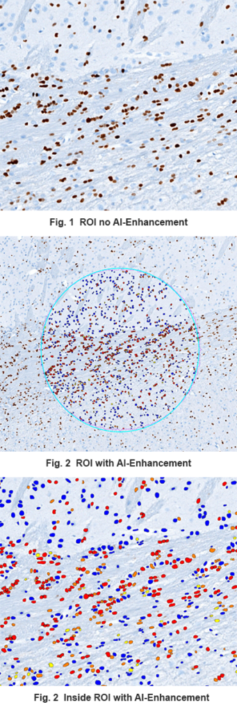
■ AI-Powered Image Analysis
Quantitative assessment of biomarkers, immune cell infiltration, and tumor microenvironment using advanced AI-driven pathology tools.
■ IHC, IF, and ISH
Advanced single and multiplex immunohistochemistry (IHC), immunofluorescence (IF), and in situ hybridization (ISH) staining for in-depth tissue profiling.
■ Whole Slide Imaging & Remote Access
Digital pathology solutions enable real-time, centralized review across global clinical sites, ensuring standardization and faster decision-making.
■ Seamless Preclinical to Clinical Integration
Our validated methodologies provide continuity in biomarker analysis, reducing variability between discovery, preclinical, and late-stage trials.
■ Fast Turnaround with Expert-Led Analysis
Optimized workflows, rapid study startup, and PhD-led teams ensure you receive high-quality, actionable data on time.
How Histology & Digital Pathology Enhance Oncology & Immunology Trials –
Supporting Immuno-Oncology & Tumor Microenvironment Analysis
Immuno-oncology (I-O) therapies rely on precise biomarker validation to assess patient response. MLM Medical Labs’ histology services provide deep insights into:
■ PD-L1, CD8, and TIL Quantification – Evaluating immune response within tumor tissue
■ Multiplex Immunofluorescence (mIF) – Capturing spatial relationships of immune cells in tumor microenvironments
■ AI-Driven Tumor Grading & Morphometric Analysis – Standardizing tumor progression assessments
Accelerating Fibrosis & Inflammatory Disease Trials
Understanding tissue remodeling, fibrosis progression, and inflammatory responses is critical in immunology trials. Our advanced histology and pathology solutions provide:
■ Fibrosis Scoring & Quantification – Using Masson’s Trichrome staining (for collagen levels) and AI-based fibrosis staging to assess tissue remodeling and disease progression.
■ Inflammation Markers (CD68, CD4, CD8) – Assessing immune cell infiltration in tissue.
■ Preclinical & Clinical Histopathology Correlation – Ensuring data consistency from early discovery to regulatory submission.
Let us know how we can help solve your most challenging tissue analysis needs.
Whether you’re conducting an immuno-oncology, fibrosis, or inflammatory disease trial, MLM Medical Labs delivers scalable, high-precision histopathology solutions to optimize your study.